Stress Fractures in Dancers

Bone tissue is one of the most metabolically active tissues in the body. It is constantly going through a process of remodelling in response the mechanical loads being placed upon it. In healthy bone, there is a homeostatic balance between bone resorption and bone formation. If this balance is disrupted for any reason, an injury to the bone can occur. In the case of a stress fracture, repetitive sub-maximal loading over time results in fatigue failure of the bone. This failure occurs along a continuum of severity with 3 distinct phases. The first phase - crack initiation - occurs at sites of stress concentration during bone loading. The second phase - crack propagation - occurs if loading continues beyond the capacity for microcrack repair. If the load continues without modification it can lead to the third phase which is a complete fracture of the bone.
The prevalence of stress fractures is between 2% - 15% in the general population. It has been found to be as high as 28% of all lower extremity injuries in patients seen in a general sports injury clinic. The annual incidence in athletes is between 1.9% - 21.1%, with distance runners reporting the highest incidence. In dancers, the lifetime incidence of stress fractures has been reported to be as high as 32%. Stress fractures also account for the greatest amount of time lost from training when compared against all other injuries in pre-professional ballet dancers.
There are well documented risk factors associated with increased rates of stress fractures. These include sex, race, nutrition (caloric insufficiency, calcium and vitamin D levels), hormonal status, low bone mineral density, BMI < 19, muscle mass/strength, neuromuscular function, abnormal bony alignment, training surfaces, improper technique/biomechanics, changes in training intensity and/or volume and overall training load. It has been found that dancers training >5hrs/day increases their risk of a stress fracture 16 times versus training <5hrs/day, regardless of other risk factors. Identifying and mitigating as many of these risk factors as possible is one key part in preventing stress fractures.
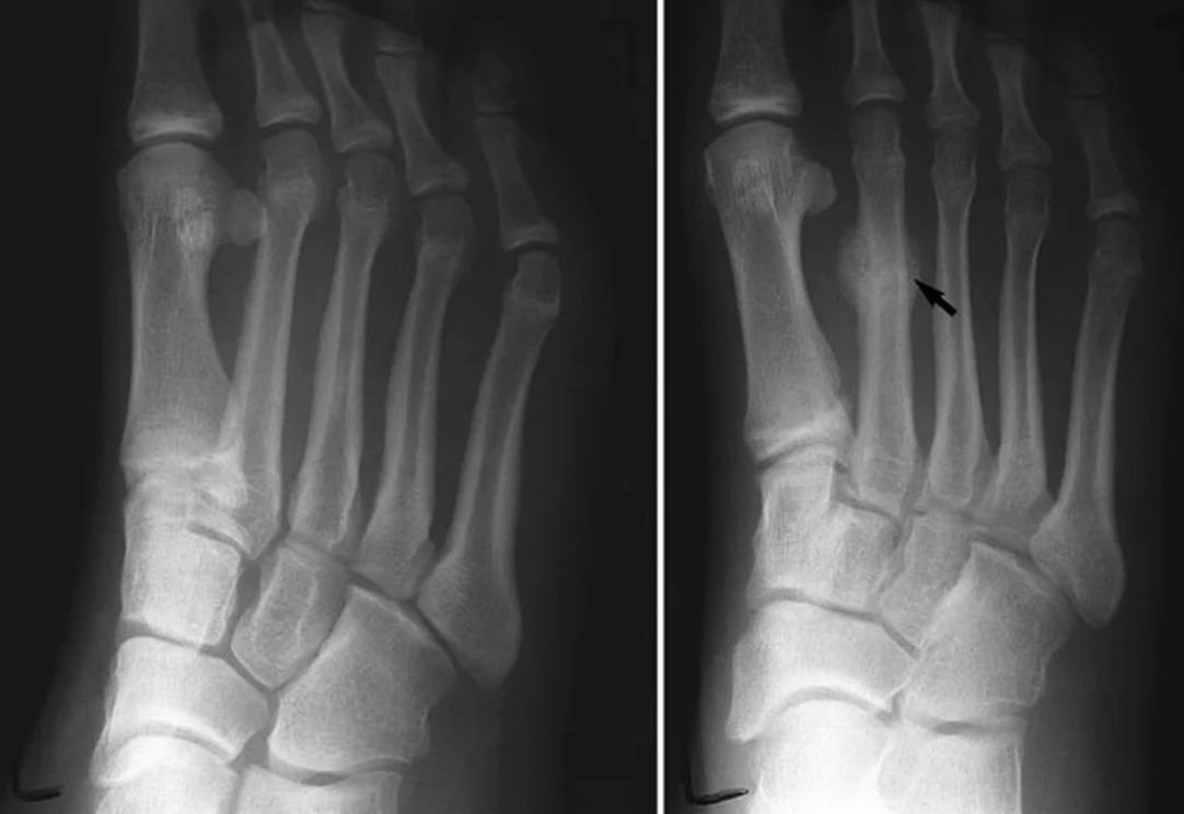
Diagnosis of a stress fracture is made by taking a detailed history, performing a physical exam and diagnostic imaging. Stress fractures will generally not be visible on a plain film x-ray, unless they are in the later stages in which case a fracture line may be visible. The gold standard for diagnosing stress fractures is an MRI. It has a sensitivity of ~100% and has the added advantage of also being able to differentiate between different bony pathology, as well as evaluate soft tissue structures at the same time. Bone scans are still commonly performed and also have a high sensitivity (~100%), however do not have the same specificity as an MRI and cannot be used to effectively monitor recovery.

Management and treatment of stress fractures in dancers depends on the risk (high vs low risk) as well as the grade of the fracture (see table 1). The risk level is determined by the anatomical location of the fracture. Low risk stress fractures that are commonly seen in dancers include the fibula, base of the 2nd metatarsal, the pars interarticularis in the spine, and the pubic ramus. Common high risk stress fractures include the shaft of the tibia, the 5th metatarsal and the sesamoid bones. Treatment will vary along a continuum from a short period of relative rest and avoiding the exacerbating activity, to possible surgical fixation of a non-union fracture.

Regardless of risk or grade of the fracture, a comprehensive treatment plan will always include identification and resolution of any associated risk factors, maintenance of fitness levels with an alternate exercise plan, rehabilitation of any associated soft tissue injuries, and a gradual return to daily activity and dance training. The gradual return to dance training should be collaboratively monitored by the health care practitioner, the dancer, and the dance teacher. Progression is based on symptom resolution and, if available, on radiographic evidence of healing. Generally a plan will progress from no/low impact activity, to low impact weight-bearing activity, to dance specific training activity and finally to unrestricted training, performance and competition. The time frame for progression will vary by dancer, anatomical site of the stress fracture and grade of the fracture. Considerations may need to be made for alternate footwear, changes in training surface and correction of any biomechanical or technique faults during this time.

In conclusion, stress fractures are a common injury seen in dancers and account for the greatest amount of time lost from training. There are many modifiable risk factors that can reduce the prevalence and incidence of stress fractures. When stress fractures do occur, a collaborative approach involving the health care team, the dancer and the dance teacher is important for a safe and effective return to dance.
References
Amanda K. Weiss Kelly and Sharon L. Hame. Managing stress fractures in athletes: the goal of diagnosis and treatment is speedy return to play. The Journal of Musculoskeletal Medicine. 27.11 (Dec. 2010): p480
Brukner P, Khan K. Clinical Sports Medicine Third Edition. Sydney Australia. McGraw-Hill Australia Ltd, 2006.
Miller TL, Best TM. Taking a Holistic approach to managing difficult stress fractures. Journal of Orthopaedic Surgery and Research. 11:98 (2016).
Fournier M. Current Concepts in lower extremity stress fractures. Current topics in sports podiatry. American Academy of Podiatric Sports Medicine. (June/July 2016): p113-118.
Ekegren CL Quested R Brodrick A. Injuries in pre-professional ballet dancers: Incidence, characteristics and consequences. Journal of Science and Medicine in Sport 17 (2014): p271-275
Brenner JS. Overuse injuries, overtraining, and burnout in child and adolescent athletes. American Academy of Paediatrics Council on Sports Medicine and Fitness. Pediatrics 119 (2007): p1242-1245
Sobhani S Dekker R Postema K Dijkstra PU. Epidemiology of foot and ankle overuse injuries in sports: A systematic review. Scand J Med Sci Sports 23 (2013): p669-686